Functional MRI does not track thoughts or electrical spikes; it measures slow hemodynamic blood-oxygen levels across 100,000 tiny 3D voxels. Without rigorous statistical corrections, fMRI produces seductive neuromyths and “voodoo correlations.”
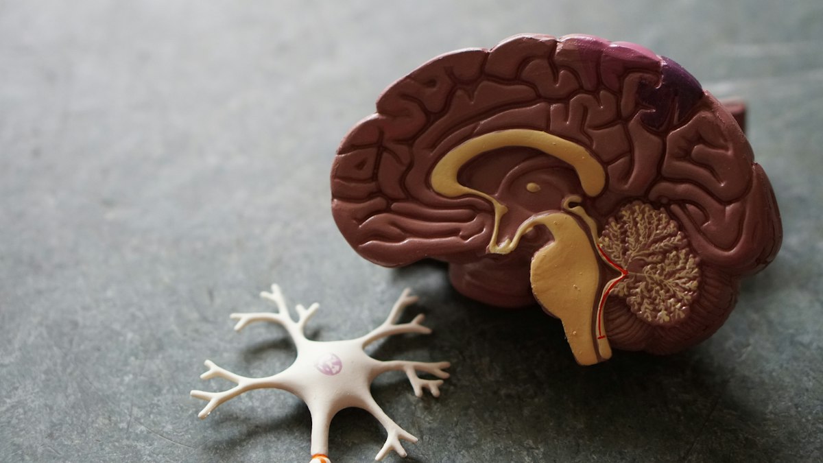
Every week, splashy media headlines announce startling discoveries about the human mind: “Scientists Find the Brain’s Jealousy Center!” “Brain Scans Reveal Why People Love Their iPhones!” “fMRI Scans Detect the God Spot in Deep Meditation!”
Accompanied by glossy 3D renderings of the human brain with glowing red, orange, and neon yellow patches, these stories carry an intoxicating aura of scientific authority. To the public, functional Magnetic Resonance Imaging (fMRI) appears like a magical window into the living soul—a direct, objective camera that photographs human thoughts, emotions, and moral intentions in real time.
In cognitive neuroscience, however, researchers view these colorful images with extreme caution. In 2009, a Dartmouth neuroscientist named Craig Bennett placed a dead Atlantic salmon inside an fMRI scanner, showed the dead fish photographs of human social situations, and asked the deceased salmon to determine what emotion the people were experiencing. Remarkably, when standard uncorrected statistical software processed the data, the dead salmon’s brain lit up with active, statistically significant neural activation!
The dead salmon experiment was a hilarious yet devastating wake-up call. To truly understand modern brain science, we must pierce the colorful veil of fMRI, uncover the physics and mathematics behind hemodynamic imaging, and learn to separate legitimate neurobiology from seductive “neuro-bunk.”
1. The Physics and Physiology of the BOLD Signal
The first fundamental truth about fMRI is that it does not measure neuronal firing. It does not measure electrical action potentials, dendritic currents, or synaptic neurotransmitter release. Instead, fMRI measures an indirect metabolic downstream consequence known as the Blood Oxygen Level Dependent (BOLD) response.
Hemodynamic Coupling and Magnetic Susceptibility
Neurons in the brain are metabolically demanding. When a specific population of neurons fires intensely, they consume glucose and oxygen. In response, local micro-capillaries dilate, rushing an over-supply of oxygenated blood to the active region—a process called neurovascular coupling.
Here is where magnetic physics enters the picture:
- Deoxygenated Hemoglobin: Has paramagnetic properties (it interacts with and distorts local magnetic fields).
- Oxygenated Hemoglobin: Has diamagnetic properties (it is magnetically neutral and does not disrupt the field).
When fresh, oxygen-rich blood floods an active brain region, it flushes away deoxygenated hemoglobin. Inside a powerful 3-Tesla superconducting magnet, this shift in magnetic susceptibility causes radiofrequency signals to decay more slowly, producing a slightly brighter image on the scan. That faint metabolic brightness is the BOLD signal.
The Temporal Sluggishness of the BOLD Response
While electrical neuronal computations occur within 1 to 10 milliseconds, the hemodynamic blood-rush is extraordinarily sluggish. The BOLD response takes roughly 2 seconds to even begin, peaks at 5 to 6 seconds, and takes up to 10 to 12 seconds to return to baseline. Trying to capture fast, real-time human thoughts with fMRI is like trying to track the speed of a racing sports car by watching the exhaust smoke drift over the highway.
2. The Mathematical Minefield: Voxels and Multiple Comparisons
An fMRI brain scan does not produce continuous photographs; it slices the brain into a 3D digital grid consisting of approximately 100,000 to 150,000 volumetric pixels (“voxels”), each measuring roughly 2 to 3 millimeters on a side. A single voxel contains up to 5.5 million neurons, tens of billions of synapses, and thousands of glia cells.
The Multiple Comparisons Nightmare
When an experimenter compares brain activity during Task A versus Task B, the computer software performs a separate statistical hypothesis test (e.g., a t-test) on every single one of those 100,000 voxels independently!
If you use a standard scientific significance threshold of p < 0.05, pure random statistical noise ensures that 5% of tests will produce false positives. In a brain with 100,000 voxels, that means pure chance will generate 5,000 falsely activated “brain centers” in every single scan! This is precisely how the dead salmon appeared to read human emotions: random thermal noise in the scanner’s magnet generated false positives across thousands of voxels.
To eliminate these ghost activations, neuroscientists must apply rigorous mathematical corrections, such as Family-Wise Error (FWE) rate correction or False Discovery Rate (FDR) algorithms. When studies fail to use rigorous corrections, bogus conclusions flood the media.
3. “Voodoo Correlations” in Social Neuroscience
In 2009, cognitive psychologist Edward Vul and colleagues dropped a bombshell paper titled “Puzzlingly High Correlations in fMRI Studies of Emotion, Personality, and Social Cognition” (originally colloquially known as the “Voodoo Correlations” paper).
Vul analyzed dozens of highly cited fMRI papers that claimed astonishingly strong statistical correlations (r > 0.85) between specific brain activations and complex psychological traits (such as empathy, racial prejudice, or neuroticism). Given the inherent measurement noise in both psychological questionnaires and fMRI scanners, such astronomical correlations are mathematically impossible.
Vul revealed that researchers were committing a cardinal statistical sin: The Circular Analysis Fallacy (Double Dipping). Researchers scanned the entire brain, selected only the voxels that showed the highest correlation with the psychological trait, and then re-calculated the correlation score on those exact same voxels! It was like predicting election winners by polling only people wearing the candidate’s campaign T-shirts.
4. The Reverse Inference Trap
Perhaps the most widespread cognitive error in neuroimaging interpretation is Reverse Inference (formalized by Russell Poldrack in 2006):
- Forward Inference (Valid): “When subjects feel fear, the amygdala activates.”
- Reverse Inference (Fallacious): “The amygdala is active in this scan, therefore the subject must be feeling fear.”
The human brain is not a collection of isolated, single-purpose hardware chips. Almost every brain region is multifunctional and involved in dozens of disparate cognitive processes. The anterior insula, for instance, activates during physical pain, romantic love, disgust, hearing unfamiliar music, reading numbers, and looking at an iPhone. Claiming that insular activation proves “love” is pure scientific fiction.
5. Modern Legitimate Neuroimaging: MVPA and Connectomics
Despite these historical methodological challenges, neuroimaging remains an indispensable, revolutionary tool when executed with modern computational rigor:
Multi-Voxel Pattern Analysis (MVPA / Neural Decoding)
Rather than asking whether a single voxel is “on” or “off,” MVPA uses machine learning classifiers to analyze complex, distributed spatial patterns of activity across thousands of voxels simultaneously. Using MVPA, researchers can decode what category of object an individual is looking at or what word they are silently reading with remarkable accuracy.
Functional Connectomics and Graph Theory
Modern neuroscience has moved away from modular “phrenology” toward Connectomics. Researchers treat the brain as a complex dynamic network of interconnected hubs, analyzing communication between the Default Mode Network, Salience Network, and Central Executive Network using sophisticated graph theory mathematics.
Frequently Asked Questions
Can fMRI read my private thoughts?
No. fMRI cannot read complex internal monologues or extract private memories. It can only classify broad categories of sensory representations or cognitive states in controlled laboratory settings where the subject cooperates fully inside a multi-million-dollar scanner.
What is the difference between structural MRI and functional MRI?
Structural MRI takes high-resolution anatomical pictures of physical brain tissue, tumors, and bones (like a 3D photograph). Functional MRI (fMRI) measures changes in blood-oxygenation over time to track metabolic activity while a subject performs cognitive tasks.
Why do neuroscientists use 3-Tesla and 7-Tesla magnets?
Tesla is a unit of magnetic field strength. A 3-Tesla scanner is roughly 60,000 times stronger than Earth’s magnetic field. Higher magnetic fields (like 7-Tesla) provide higher signal-to-noise ratios, allowing researchers to resolve smaller voxels and inspect finer cortical layers.
Are EEG and fMRI complementary?
Yes. EEG has superb millisecond temporal resolution but poor spatial resolution. fMRI has superb millimeter spatial resolution but sluggish temporal resolution. Combining both provides a comprehensive view of brain function.
Can fMRI be used as a lie detector in court?
Federal and international courts overwhelmingly reject fMRI lie detection as scientifically unreliable. Variations in anxiety, memory recall, and deliberate mental counter-measures (such as wiggling toes or doing mental arithmetic) easily distort BOLD activations.
fMRI is not mind-reading; it is complex statistical modeling of biological fluid dynamics. When stripped of sensationalism and held to rigorous mathematical standards, neuroimaging illuminates the sublime architecture of the human mind without reducing the soul to a colorful cartoon.
